 Explore
 Validate
 Learn
Learn Western blot
 ELISA
ELISAAntibody data
- Antibody Data
- Antigen structure
- References [0]
- Comments [0]
- Validations
- Western blot [2]
- Immunohistochemistry [5]
- Flow cytometry [2]
Submit
Validation data
Reference
Comment
Report error
- Product number
- NBP2-34675-0.1 mg - Provider product page

- Provider
- Novus Biologicals
- Product name
- Mouse Monoclonal Cytokeratin 14 Antibody
- Antibody type
- Monoclonal
- Description
- Protein A purified. Cytokeratin 14 (CK14) belongs to the type I (or A or acidic) subfamily of low molecular weight keratins and exists in combination with keratin 5 (type II or B or basic). CK14 is found in basal cells of squamous epithelia, some glandular epithelia, myoepithelium, and mesothelial cells. Anti-CK14 is useful in differentiating squamous cell carcinomas from poorly differentiated epithelial tumors. Anti-CK14 is one of the specific basal markers for distinguishing between basal and non-basal subtypes of breast carcinomas. Anti-CK14 is also a good marker for differentiation of intraductal from invasive salivary duct carcinoma by the positive staining of basal cells surrounding the in-situ neoplasm as well as for differentiation of benign prostate from prostate carcinoma. Furthermore, this antibody has been useful in separating oncocytic tumors of the kidney from its renal mimics, and in identifying metaplastic carcinomas of the breast.
- Reactivity
- Human, Mouse, Rat
- Host
- Mouse
- Isotype
- IgG
- Vial size
- 0.1 mg
- Concentration
- 1.0 mg/ml
- Storage
- Store at 4C short term. Aliquot and store at -20C long term. Avoid freeze-thaw cycles.
No comments: Submit comment
Supportive validation
- Submitted by
- Novus Biologicals (provider)
- Main image

- Experimental details
- Simple Western: Cytokeratin 14 Antibody (LL002) - Azide and BSA Free [NBP2-34675] - Simple Western lane view shows a specific band for Cytokeratin 14 in 0.2 mg/ml of A431 lysate(s). This experiment was performed under reducing conditions using the 12-230 kDa separation system.

- Submitted by
- Novus Biologicals (provider)
- Main image

- Experimental details
- Simple Western: Cytokeratin 14 Antibody (LL002) - Azide and BSA Free [NBP2-34675] - Electropherogram image of the corresponding Simple Western lane. Cytokeratin 14 antibody was used at 10 ug/ml dilution of A431 lysates(s) respectively.

Supportive validation
- Submitted by
- Novus Biologicals (provider)
- Main image

- Experimental details
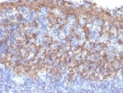
- Immunohistochemistry-Paraffin: Cytokeratin 14 Antibody (LL002) - Azide and BSA Free [NBP2-34675] - Formalin-fixed, paraffin-embedded prostate stained with Cytokeratin 14 Monoclonal Antibody (LL002).

- Submitted by
- Novus Biologicals (provider)
- Main image

- Experimental details
- Immunohistochemistry-Paraffin: Cytokeratin 14 Antibody (LL002) - Azide and BSA Free [NBP2-34675] - Formalin-fixed, paraffin-embedded human prostate (20X) stained with Cytokeratin 14 MAb (LL002).

- Submitted by
- Novus Biologicals (provider)
- Main image

- Experimental details
- Immunohistochemistry-Paraffin: Cytokeratin 14 Antibody (LL002) - Azide and BSA Free [NBP2-34675] - Formalin-fixed, paraffin-embedded human Prostate Carcinoma stained with Cytokeratin 14 Antibody (LL002).

- Submitted by
- Novus Biologicals (provider)
- Main image
- Experimental details
- Immunohistochemistry-Paraffin: Cytokeratin 14 Antibody (LL002) - Azide and BSA Free [NBP2-34675] - Formalin-fixed, paraffin-embedded human Prostate stained with Cytokeratin 14 Monoclonal Antibody (LL002)

- Submitted by
- Novus Biologicals (provider)
- Main image

- Experimental details
- Immunohistochemistry-Paraffin: Cytokeratin 14 Antibody (LL002) - Azide and BSA Free [NBP2-34675] - Formalin-fixed, paraffin-embedded human prostate (10X) stained with Cytokeratin 14 MAb (LL002).

Supportive validation
- Submitted by
- Novus Biologicals (provider)
- Main image
- Experimental details
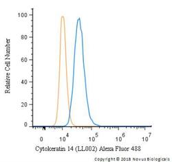
- Flow Cytometry: Cytokeratin 14 Antibody (LL002) - Azide and BSA Free [NBP2-34675] - An intracellular stain was performed on HeLa cells with Cytokeratin 14 Antibody (LL002) NBP2-34675AF488 (blue) and a matched isotype control (orange). Cells were fixed with 4% PFA and then permeablized with 0.1% saponin. Cells were incubated in an antibody dilution of 10 ug/mL for 30 minutes. Both antibodies were conjugated to Alexa Fluor 488.

- Submitted by
- Novus Biologicals (provider)
- Main image

- Experimental details
- Flow Cytometry: Cytokeratin 14 Antibody (LL002) - Azide and BSA Free [NBP2-34675] - Flow Cytometric Analysis of trypsinized MeOH-fixed HeLa cells using Cytokeratin 14 Antibody (LL002) followed by Goat anti-Mouse IgG-CF488 (Blue); Isotype Control (Red).
